AUTORAS
Mariane Fontana Mezoni
Graduanda em Odontologia – Centro Universitário Unisep.
Orcid: 0000-0002-4329-3152.
Marta Aparecida Alberton Nuernberg
Professora doutora do curso de Odontologia – Centro Universitário Unisep.
Orcid: 0000-0003-2167-9772.
RESUMO
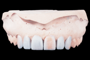
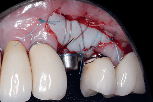
A manutenção dos tecidos periodontais em torno dos implantes tornou-se uma medida indispensável para assegurar a saúde e a estética esperadas ao final da reabilitação. Estudos têm relacionado a quantidade adequada de mucosa queratinizada como fator de proteção e resistência contra traumas mecânicos e biológicos à mucosa peri-implantar. Dessa forma, o presente trabalho teve como objetivo relatar um caso clínico de aumento de tecido queratinizado (TQ) em área peri-implantar utilizando enxerto gengival livre (EGL). Paciente do sexo feminino com 57 anos de idade, ASA II, tinha um implante dentário instalado há seis meses na região do 24. No exame clínico, constatou-se uma espessura inadequada de TQ, assim como fenótipo periodontal fino na área peri-implantar. Levando em consideração as características clínicas do caso, foi indicada a modificação de fenótipo gengival peri-implantar por meio de EGL. A escolha da abordagem cirúrgica foi direcionada pela fase de condicionamento gengival com cicatrizador em que a paciente se encontrava. O enxerto foi obtido do palato. No acompanhamento pós-operatório de 30 dias, foi observado um ganho de tecido queratinizado > 2 mm com proteção completa da área peri-implantar e relato de maior conforto durante a higienização. A técnica de EGL empregada demonstrou resultados satisfatórios, com ganho expressivo de TQ, além da consequente melhora na estabilidade tecidual, segurança e saúde peri-implantar.
Palavras-chave – Implantes dentários; Placa bacteriana; Fenótipo; Enxerto de tecidos.
ABSTRACT
The maintenance of peri-implant soft tissues has become an essential measure to ensure the health and esthetics expected at the end of implant rehabilitation. Studies have related the adequate amount of keratinized mucosa as a protection and resistance factor against mechanical and biological trauma to the peri-implant mucosa. Thus, the present study aimed to report a clinical case of increase in keratinized tissue (KT) in the peri-implant area using free gingival graft (FGG). A 57-year-old female patient, with dental implant installed 6 months ago in the 24 region. The clinical examination revealed an inadequate thickness of KT, as well as a thin periodontal phenotype in the peri-implant area. Considering the clinical characteristics of the case, the modification of the periimplant gingival phenotype through FGG was indicated. The choice of surgical approach was guided by the gingival conditioning phase with the healing abutment. The graft was taken from the palate. In the 30-day postoperative follow-up, a gain of > 2 mm of KT was observed, with complete protection of the peri-implant area and a report of greater comfort during cleaning. The FGG technique employed showed satisfactory results, with expressive KT gain, in addition to the consequent improvement in tissue stability, safety and peri-implant health.
Key words – Dental implants; Dental plaque; Phenotype; Tissue transplantation.
Referências
- Wang Y, Zhang Y, Miron RJ. Health, maintenance, and recovery of soft tissues around implants. Clin Implant Dent Relat Res 2016;18(3):618-34.
- Chackartchi T, Romanos GE, Sculean A. Soft tissue-related complications and management around dental implants. Periodontol 2000 2019;81(1):124-38.
- Golmayo P, Barallat L, Losada M, Valles C, Nart J, Pascual-La Rocca A. Keratinized tissue gain after free gingival graft augmentation procedures around teeth and dental implants: a prospective observational study. J Clin Periodontol 2021;48(2):302-14.
- Tavelli L, Barootchi S, Avila-Ortiz G, Urban IA, Giannobile WV, Wang HL. Peri-implant soft tissue phenotype modification and its impact on peri-implant health: a systematic review and network meta-analysis. J Periodontol 2021;92(1):21-44.
- Malpartida-Carrillo V, Tinedo-Lopez PL, Guerrero ME, Amaya-Pajares SP, Özcan M, Rösing CK. Periodontal phenotype: a review of historical and current classifications evaluating different methods and characteristics. J Esthet Restor Dent 2021;33(3):432-45.
- Avila-Ortiz G, Gonzalez-Martin O, Couso-Queiruga E, Wang HL. The peri-implant phenotype. J Periodontol 2020;91(3):283-8.
- Brito C, Tenenbaum HC, Wong BK, Schmitt C, Nogueira-Filho G. Is keratinized mucosa indispensable to maintain peri-implant health? A systematic review of the literature. J Biomed Mater Res B Appl Biomater 2014;102(3):643-50.
- Raoofi S, Asadinejad SM, Khorshidi H. Evaluation of color and width of attached gingiva gain in two surgical techniques: free gingival graft and connective tissue graft covered by thin mucosal flap, a clinical trial. J Dent (Shiraz) 2019;20(4):224-31.
- Almeida JM, Novaes VN, Faleiros PL, Macarimi VC, Bosco AF, Theodoro LH et al. Increased keratinized gingiva in peri-implant mucosa. Revista de Odontologia da Unesp 2012;41(5):365-9.
- Agudio G, Chambrone L, Selvaggi F, Pini-Prato GP. Effect of gingival augmentation procedure (free gingival graft) on reducing the risk of non-carious cervical lesions: a 25- to 30-year follow-up study. J Periodontol 2019;90(11):1235-43.
- Lang NP, Berglundh T, Working Group 4 of Seventh European Workshop on Periodontology. Periimplant diseases: where are we now? – Consensus of the Seventh European Workshop on Periodontology. J Clin Periodontol 2011;38(suppl.11):178-81.
- Akcan SK, Güler B, Hatipoğlu H. The effect of different gingival phenotypes on dimensional stability of free gingival graft: a comparative 6-month clinical study. J Periodontol 2019;90(7):709-17.
- Zucchelli G, Tavelli L, McGuire MK, Rasperini G, Feinberg SE, Wang HL et al. Autogenous soft tissue grafting for periodontal and peri-implant plastic surgical reconstruction. J Periodontol 2020;91(1):9-16.
- Cairo F, Pagliaro U, Nieri M. Soft tissue management at implant sites. J Clin Periodontol 2008;35(8 suppl.):163-7.