AUTORES
Karine Ferreira Teixeira
Especialista em Periodontia e mestranda em Ciências Odontológicas Aplicadas (área de Periodontia) – FOB-USP. Orcid: 0000-0001-6588-2233.
Amanda de Oliveira Macedo
Especialista em Periodontia e mestranda em Ciências Odontológicas Aplicadas (área de Periodontia) – FOB-USP. Orcid: 0000-0001-5991-4585.
Andréia Pereira de Souza-Pavani
Doutora em Periodontia e mestra em Ciências Odontológicas Aplicadas (área de Reabilitação Oral, com linha de pesquisa em Periodontia) – FOB-USP. Orcid: 0000-0003-2735-512X.
Carla Andreotti Damante
Professora doutora associada da disciplina de Periodontia – FOB-USP. Orcid: 0000-0002-6782-8596.
Adriana Campos Passanezi Sant’Ana
Professora doutora associada da disciplina de Periodontia – FOB-USP. Orcid: 0000-0001-5640-9292.
Mariana Schutzer Ragghianti Zangrando
Professora doutora associada da disciplina de Periodontia – FOB-USP. Orcid: 0000-0003-0286-7575.
RESUMO
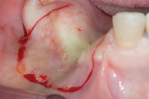
O planejamento cirúrgico adjunto às técnicas de cirurgia plástica periodontal com as biopsias excisionais de lesões gengivais exofíticas (de origem reacional ou neoplásicas) pode minimizar os defeitos mucogengivais inerentes à remoção da lesão. Este caso clínico descreveu a remoção de um fibroma odontogênico periférico (FOP), um tumor odontogênico benigno, na região posterior de maxila por biopsia excisional e com controle pós-operatório de 16 meses. Um retalho pediculado com divisão mista e deslizado lateralmente foi associado visando minimizar as sequelas da remoção da lesão, promovendo adequado fechamento do defeito. A conduta associativa da biopsia excisional ao deslize lateral de retalho demonstrou ser efetiva, já que não houve recidiva da lesão, e proporcionou manutenção da arquitetura periodontal. Portanto, o planejamento periodontal prévio de abordagens em lesões gengivais é importante para a manutenção da função e estética periodontal. Além disso, a associação dos procedimentos cirúrgicos de plástica periodontal e biopsia excisional em uma única etapa demonstrou resultados estéticos satisfatórios e preservação da faixa de mucosa ceratinizada.
Palavras-chave – Biopsia; Crescimento excessivo da gengiva; Tumores odontogênicos; Cirurgia mucogengival; Relato de caso.
ABSTRACT
Surgical planning of periodontal plastic surgery techniques with excisional biopsies of exophytic gingival lesions (of reactional or neoplastic origin) can minimize mucogingival defects inherent in the removal of the lesion. This clinical case describes the removal of a benign peripheral odontogenic fibroma (POF) in the posterior region of the maxilla by excisional biopsy and a 16-month postoperative control. A laterally positioned pediculated flap with mixed division was designed to minimize the sequelae of lesion removal, promoting adequate closure of the defect. The association of the excisional biopsy to the laterally positioned flap proved to be effective since there was no recurrence of the lesion and maintenance of the periodontal architecture was provided. Therefore, prior periodontal planning of approaches in gingival lesions is important for the maintenance of periodontal function and aesthetics. In addition, the association of periodontal plastic surgical procedures and excisional biopsy in a single step demonstrated satisfactory aesthetic results and preservation of the keratinized mucosa width.
Key words – Biopsy; Gingival overgrowth; Odontogenic tumors; Mucogingival surgery; Case report.
Referências
- Miller Jr. PD. Root coverage grafting for regeneration and aesthetics. Periodontol 2000 1993;1(1):118-27.
- Miller Jr. PD. Regenerative and reconstructive periodontal plastic surgery. Mucogingival surgery. Dent Clin North Am 1988;32(2):287-306.
- de Matos FR, Benevenuto TG, Nonaka CFW, Pinto LP, de Souza LB. Retrospective analysis of the histopathologic features of 288 cases of reactional lesions in gingiva and alveolar ridge. Appl Immunohistochem Mol Morphol 2014;22(7):505-10.
- Buchner A, Shnaiderman-Shapiro A, Vered M. Relative frequency of localized reactive hyperplastic lesions of the gingiva: a retrospective study of 1675 cases from Israel. J Oral Pathol Med 2010;39(8):631-8.
- Henriques PS, Okajima LS, Nunes MP, Montalli VA. Coverage root after removing peripheral ossifying fibroma: 5-year follow-up case report. Case Rep Dent 2016;2016:6874235.
- Choudary SA, Naik AR, Naik MS, Anvitha D. Multicentric variant of peripheral ossifying fibroma. Indian J Dent Res 2014;25(2):220-4.
- Bosco AF, Bonfante S, Luize DS, Bosco JM, Garcia VG. Periodontal plastic surgery associated with treatment for the removal of gingival overgrowth. J Periodontol 2006;77(5):922-8.
- Vogan WI. Immediate repair of gingival biopsy sites. Oral Surg Oral Med Oral Pathol 1975;40(3):333-5.
- Passanezi E, Sant’Ana ACP, Rezende MLR, Greghi SLA, Janson WA. Distâncias biológicas periodontais princípios para a reconstrução periodontal, estética e protética (1a). São Paulo: Artes Médicas, 2011. p.304.
- Capelozza ALA, Moreira CR, Ferraz BFR, Sant’ana LFM, Lara VS. Fibroma odontogênico periférico: revisão da literatura e relato de caso. Arq Odontol 2016;43(1).
- Chan JKC, El-Naggar AK, Grandis JR, Takata T, Slootweg PJ. WHO Classification of Head and Neck Tumours (4th ed.). World Health Organization 2017. p.347.
- Wright JM, Vered M. Update from the 4th edition of the World Health Organization classification of head and neck tumours: odontogenic and maxillofacial bone tumors. Head Neck Pathol 2017;11(1):68-77.
- Tolentino E. Nova classificação da OMS para tumores odontogênicos: o que mudou? [On-line]. Disponível em <http://seer.upf.br/index.php/rfo/article/view/7905>. Acesso em: 7-12-2021.
- Neville BW, Damm DD, Allen CM, Bouquot JE. Patologia Oral & Maxilofacial (3a). Rio de Janeiro: Guanabara Koogan, 2009. p.992.
- Kenney JN, Kaugars GE, Abbey LM. Comparison between the peripheral ossifying fibroma and peripheral odontogenic fibroma. J Oral Maxillofac Surg 1989;47(4):378-82.
- Slabbert HV, Altini M. Peripheral odontogenic fibroma: a clinicopathologic study. Oral Surg Oral Med Oral Pathol 1991;72(1):86-90.
- Manor Y, Mardinger O, Katz J, Taicher S, Hirshberg A. Peripheral odontogenic tumours – differential diagnosis in gingival lesions. Int J Oral Maxillofac Surg 2004;33(3):268-73.
- de-Sena LSB, Miguel MCC, Pereira JV, Gomes DQC, Alves PM, Nonaka CFW. Fibroma odontogênico periférico em gengiva mandibular: relato de caso. J Bras Patol Med Lab 2019;55(2):192-201.
- Buchner A, Merrell PW, Carpenter WM. Relative frequency of peripheral odontogenic tumors: a study of 45 new cases and comparison with studies from the literature. J Oral Pathol Med 2006;35(7):385-91.
- Silva LP, Macedo RAP, Serpa MS, Sobral APV, Souza LB. Global frequency of benign and malignant odontogenic tumors according to the 2005 WHO classification. J Oral Diag 2017;2(1).
- Cairo F. Periodontal plastic surgery of gingival recessions at single and multiple teeth. Periodontol 2000 2017;75(1):296-316.
- Group H, Warren R. Repair of gingival defects by a sliding flap operation. J Periodontol 1956;27(2):92-5.
- Ruben MP, Goldman HM, Janson W. Biological considerations fundamental to successful employment of laterally repositioned pedicle flaps and free autogenous gingival grafts in periodontal therapy. In: Stahl S, editor. Periodontal Surgery. Springfield: Illinois, 1976.


