AUTOR
Paulo Henrique Orlato Rossetti
Cirurgião-dentista, mestre e doutor em Reabilitação Oral – FOB-USP. Orcid: 0000-0002-0868-6022.
RESUMO
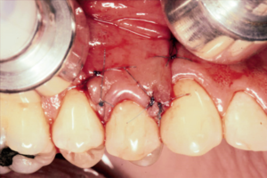
Objetivo: revisitar os momentos que nos trouxeram às condutas clínicas sobre o gerenciamento do tecido gengival na extração/ colocação imediata de implantes dentários unitários. Material e métodos: uma busca foi realizada em mais de 20 periódicos científicos, acessados número por número, apenas no ano de 2003. Resultados: três artigos foram escolhidos como os mais representativos deste tópico. O primeiro traz os resultados clínicos um ano depois da extração e colocação/provisionalização imediata do implante na zona estética: as variações nas margens gengivais mesial (de 0,31 mm para 0,53 mm) e vestibulomediana (de 0,36 mm para 0,55 mm) são similares e levemente diferentes das variações nas margens gengivais distais (de 0,21 mm para 0,39 mm), com variações nos níveis ósseos mesiais e distais de 0,26 e 0,28 mm, dentro do esperado. O segundo mostrou o que aconteceria se dois implantes dentários adjacentes fossem colocados em tempos diferentes, respeitando a osseointegração: aqui, o índice de papila variou de 3 para 2,75, onde mais uma vez os índices ósseos marginais se mostraram abaixo de 1 mm de variação. Finalmente, o terceiro avaliou as dimensões da mucosa peri-implantar de implantes de dois estágios, constatando que, mesmo havendo um nível de sondagem maior nas faces mesial e distal peri-implantar, as faces dentárias proximais aos implantes permanecem estáveis, fornecendo índices similares aos observados em dentes naturais. Isto mostrou que o nível da papila peri-implantar depende apenas do nível ósseo do dente vizinho. Conclusão: variações nas papilas interdentárias de elementos extraídos na região estética são suavizadas pela seleção adequada dos pacientes, protocolo cirúrgico e customização dos componentes protéticos.
Palavras-chave – Tecido gengival; Papila dentária; Implante dentário; Extração dentária.
ABSTRACT
Objective: to revisit the moments that brought us to the clinical management of the gingival tissue upon single-tooth removal/ immediate implant placement. Material and methods: a search was conducted in more than 20 scientific periodicals, accessed number by number, only at the year 2003. Results: three articles were selected as the most representative of this topic: the first brought the clinical outcomes one year later after tooth extraction and immediate placement/provisionalization at the esthetic zone: the variations at the mesial (0.31 mm to 0.53 mm) and facial (0.36 mm to 0.55 mm) gingival margins were very similar and slightly different from the distal gingival margins (0.21 mm to 0.39 mm), with changes at the mesial and distal bone levels of 0.26 mm and 0.28 mm, respectively and within normal ranges. The second showed what happened to adjacent dental implants placed at different times respecting the osseointegration periods: for this, the papilla index score changed from 3 to 2.75. Once again, the marginal bone levels remained below 1 mm. Finally, the third article evaluated the dimensions of the peri-implant mucosa for 2-stage dental implants, highlighting that even when the bone sounding was greater at the mesial and distal peri-implant aspects, the proximal tooth aspects remained stable providing similar indexes to that seen for natural neighborhood teeth, show that peri-implant papillae level relies on the bone level of adjacent tooth. Conclusion: changes at the interdental papillae for extracted tooth in the esthetic zone are alleviated by adequate patient selection, surgical protocol, and customization of prosthetic components.
Key words – Gingival tissue; Dental papilla; Dental implant; Tooth extraction.
Referências
- Kois JC. Predicatable single tooth esthetics. The five diagnostic keys. Compend Contin Dent Educ 2001;22(3):199-206.
- Schroeder HE. Structure of gingiva. In: Schroeder HE (ed.). The periodontium. Berlin: Springer Verlag, 1986. p.239-300.
- Zetu L, Wang HL. Management of the inter-dental/inter-implant papilla. J Clin Periodontol 2005;32(7):831-9.
- Shonohara Gonzalez MK, Almeida ALPF, Greghi SLA, Pegoraro LF, Mondelli J, Moreno T. Interdental papillary house: a new guide and concept for clinicians. Int J Periodontics Restorative Dent 2011;31(6):e87-e93.
- Samet N, Jotkowitz A. Classification and prognosis evaluation of individual teeth: a comprehensive approach. Quintessence Int 2009;40(5):377-87.
- Kan JYK, Rungcharassaeng K. Immediate placement and provisionalization of maxillary anterior single tooth implants: a surgical and prosthodontic rationale. Pract Periodont Aesthet Dent 2000;12(9):817-24.
- Kan JYK, Rungcharassaeng K, Lozada J. Immediate placement and provisionalization of maxillary anterior single implants: 1-year prospective study. Int J Oral Maxillofac Implants 2003;18(1):31-9.
- Kan JYK, Rungcharassaeng K. Interimplant papilla preservation in the esthetic zone: a report of six consecutive cases. Int J Periodontics Restorative Dent 2003;23(3):249-59.
- Jemt T. Regeneration of gingival papillae after single implant treatment. Int J Periodontics Restorative Dent 1997;17(4):326-33.
- Kan JYK, Rungcharassaeng K, Umezu K, Kois JC. Dimensions of peri-implant mucosa: an evaluation of maxillary anterior single implants in humans. J Periodontol 2003;74(4):557-62.
- Rossetti PHO. Papilas e implantes dentários: a última fronteira. ImplantNewsPerio 2016;1(2):250-4.
- Nobel Replace Select System [On-line]. Disponível em <https://store.nobelbiocare.com/us/en/implants/nobelreplace-select-tapered/replace-select-tapered)>.
- Pow EH, McMillian AS. A modified implant healing abutment to optimize soft tissue contours: a case report. Implant Dent 2004;13(4):297-300.
- Tetelman EV, Babbush CA. A new transitional abutment for immediate aesthetics and function. Implant Dent 2008;17(1):51-8.
- Gallucci GO, Belser UC, Bernard JP, Magne P. Modeling and characterization of the CEJ for optimization of the esthetic implant design. Int J Periodontics Restorative Dent 2004;24(1):19-29.
- Kan JYK, Rungcharassaeng K, Lozada JL, Zimmermann G. Facial gingival tissue stability following immediate placement and provisionalization of maxillary anterior single implants: a 2- to 8-year follow-up. Int J Oral Maxillofac Implants 2011;26(1):179-87.
- Lewgoy HR, Matson R, Rufaiel M, Matsushita MM, Forger SI, Tortamano P et al. Estabelecimento de um protocolo de manutenção para prevenção de mucosites e peri-implantites. ImplantNews 2012;9(1):11-9.