AUTORES
Leandro Miranda Ribeiro Dias
Mestrando em Prótese e Reabilitação Oral – SLMandic; Especialista em Bucomaxilofacial – FOA; Especialista em Implantodontia Oral – Unigranrio. Orcid: 0000-0002-7230-2142.
Milton Edson Miranda
Doutor em Prótese Dentária – Universidade de São Paulo; Professor do curso de pós-graduação – SLMandic. Orcid: 0000-0002-5410-6500.
Rafael Pino Vitti
Doutor em Materiais Dentários – FOP-Unicamp; Professor do curso de Odontologia – Centro Universitário da Fundação Hermínio Ometto. Orcid: 0000-0001-6366-5868.
William Cunha Brandt
Doutor em Materiais Dentários e Aperfeiçoamento em Endodontia Clínica – FOP-Unicamp; Professor da pós-graduação em Implantodontia – Unisa. Orcid: 0000-0002-6362-0499.
RESUMO
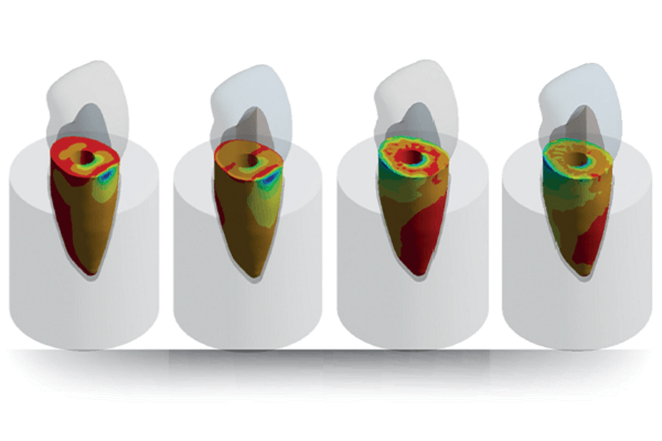
Objetivo: no presente estudo in silico foi avaliar a distribuição de tensões promovidas por um movimento em um incisivo central superior restaurado com pinos intrarradiculares feitos em polieteretercetona (PEEK) e pino de fibra de vidro (PFV) com coroa de dissilicato de lítio. Material e métodos: através de uma tomografia, criou-se modelos tridimensionais para análise de elementos finitos e reproduziu-se a região anterior da maxila, constituída em sua densidade por tecido ósseo do tipo três. Utilizou-se em cada modelo um elemento 11 com uma raiz com 13 mm de comprimento e uma coroa com 10 mm de altura. Foram obtidos dois grupos experimentais modelados com o auxílio de um software, que testaram as tensões máxima principal e mínima principal em dois tipos de pinos (PFV e PEEK) com 9 mm de altura, e aplicada força na face palatina de 100 N em quatro carregamentos de movimentos: cervical, médio, incisal e axial. Para a análise biomecânica, utilizou-se um software de análise de elementos finitos. Os resultados foram obtidos e avaliados por meio das tensões máxima principal (tração) e mínima principal (compressão) na raiz do elemento dentário. Resultados: o PEEK apresentou valores de tensão máxima principal maiores do que o PFV em todos os movimentos. Já na tensão mínima principal, o PEEK apresentou maiores valores que o PFV nos movimentos cervical e incisal, e o PFV apresentou maiores valores que o PEEK nos movimentos médio e axial. Conclusão: o PFV mostrou melhor destribuição de tensões na raiz do dente, quando comparado ao PEEK.
Palavras-chave – PEEK; Pino termoplástico sintético; Pino fibra de vidro.
ABSTRACT
Objective: to evaluate, in silico, the stress distribution promoted by movement in a maxillary central incisor restored with intraradicular posts made of polyetheretherketone (PEEK) and fiberglass post (FGP) with lithium disilicate crown. Material and methods: a CBCT was used to create three-dimensional models for finite element analysis, reproducing the anterior region of the maxilla, constituted in its density by type three bone tissue. An element 11 (crown height: 10 mm; rooth length:13 mm) was used in each model. Two experimental groups were modeled with the aid of software were obtained, which tested the maximum principal and minimum principal stresses on two post types (FGP and PEEK) with 9 mm in height and applied a force of 100 N on the palatal surface in four movement loads: cervical, medium, incisal and axial. Biomechanical analysis was provided with the aid of a dedicated software. The results were obtained and evaluated by means of the maximum principal stress (tension) and minimum principal stress (compression) in the root of the dental element. Results: PEEK presented the highest principal maximum stress values than FGP in all movements. In the minimum principal tension, PEEK presented the highest values than FGP in the cervical and incisal movements and FGP presented the highest values than PEEK in the medium and axial movements. Conclusion: it can be concluded that FGP showed better stress distribution in the tooth root than PEEK.
Key words – PEEK; Synthetic thermoplastic post; Fiberglass post.
Referências
- Ibrahim RO, Al-Zahawi AR, Sabri LA. Mechanical and thermal stress evaluation of PEEK prefabricated post with different head design in endodontically treated tooth: 3D-finite element analysis. Dent Mater J 2021;40(2):508-18.
- Lee K-S, Shin J-H, Kim J-E, Kim J-H, Lee W-C, Shin S-W et al. Biomechanical evaluation of a tooth restored with high performance polymer PEKK post-core system: a 3d finite element analysis. BioMed Research International 2017;2017:1373127.
- Gan K, Liu H, Liu X, Niu D. Research progress of polyether ether ketone biocomposites. Ann Materials Sci Eng 2015;2(1):1020.
- Najeeb S, Zafar MS, Khurshid Z, Siddiqui F. Applications of polyetheretherketone (PEEK) in oral implantology and prosthodontics. J Prosthodont Res 2016;60(1):12-9.
- Rocha RF, Anami LC, Campos TM, Melo RM, Souza RO, Bottino MA. Bonding of the polymer polyetheretherketone (PEEK) to human dentin: effect of surface treatments. Braz Dent J 2016;27(6):693-9.
- Song CH, Choi JW, Jeon YC, Jeong CM, Lee SH, Kang ES et al. Comparison of the microtensile bond strength of a polyetheretherketone (PEEK) tooth post cemented with various surface treatments and various resin cements. Materials (Basel) 2018;11(6):916.
- Skinner HB. Composite technology for total hip arthroplasty. Clin Orthop Relat Res 1988;(235):224-36.
- Katzer A, Marquardt H, Westendorf J, Wening JV, von Foerster G. Polyetheretherketone – cytotoxicity and mutagenicity in vitro. Biomaterials 2002;23(8):1749-59.
- Novis RM, Cardoso MCP, Ribeiro FC, Silva EVF, León BLT. Avaliação da resistência ao cisalhamento do pino pré-fabricado pelo teste push-out, utilizando dois sistemas cimentantes autocondicionantes. Rev Odontol Araçatuba 2013;34(1):39-44.
- Pegoraro LF, do Valle AL, Araújo CRP, Bonfante G, Conti PCR. Prótese fixa: bases para o planejamento em reabilitação oral (2a). São Paulo: Artes Médicas, 2013.
- Pereira JR, Rosa RA, Só MVR, Afonso D, Kuga MC, Honório HM et al. Push-out bond strength of fiber posts to root dentin using glass ionomer and resin modified glass ionomer cements. J Appl Oral Sci 2014;22(5):390-6.
- Filho FJS, Pacheco RR, Caiado ACRL. Endodontia passo a passo: evidências clínicas. São Paulo: Artes Médicas, 2015.
- Andrioli ARV, Coutinho M, Vasconcellos AA, Miranda ME. Relining effects on the push-out shear bond strength of glass fiber posts. Rev Odontol Unesp 2016;45(4):227-33.
- Çaglar A, Bal BT, Karakoca S, Aydın C, Yılmaz H, Sarısoy S. Three-dimensional finite element analysis of titanium and yttrium-stabilized zirconium dioxide abutments and implants. Int J Oral Maxillofac Implants 2011;26(5):961-9.
- Ko CC, Chu CS, Chung KH, Lee MC. Effects of posts on dentin stress distribution in pulpless teeth. J Prosthet Dent 1992;68(3):421-7.
- Ereifej N, Rodrigues FP, Silikas N, Watts DC. Experimental and FE shear-bonding strength at core/veneer interfaces in bilayered ceramics. Dent Mater 2011;27(6):590-7.
- Ferreira MBDC et al. Pino de fibra de vidro anatômico: relato de caso. Journal of Oral Investigations 2018;7(1):52-61.
- Lee KS, Shin JH, Kim JE, Kim JH, Lee WC, Shin SW et al. Biomechanical assessment of a tooth restored with a high performance polymer PEEK post-core system: a 3d finite element analysis. BioMed Res Int 2017;1373127:1-9.
- Lee WT, Koak JY, Lim YJ, Kim SK, Kwon HB, Kim MJ. Stress shielding and fatigue limits of poly-ether-ether-ketone dental implants. J Biomed Mater Res Part B 2012;100(4):1044-52.
- Bathala L, Majeti V, Rachuri N, Singh N, Gedela S. The role of polyether ether ketone (Peek) in Dentistry – a review. J Med Life 2019;12(1):5-9.
- Soares NS, Sant´ana LLP. Estudo comparativo entre pino de fibra de vidro e pino metálico fundido: uma revisão de literatura. Id On Line Rev Mult Psic 2018;12(42):996-1005.
- Monich PR, Berti FV, Porto LM, Henriques B, Oliveira APN, Fredel MC et al. Avaliação físico-química e biológica de compósitos de PEEK incorporando fibras naturais de sílica amorfa para aplicações biomédicas. Cienc Eng Mat 2017;79:354-62.
- Rahmitasari F, Ishida Y, Kurahashi K, Matsuda T, Watanabe M, Ichikawa T. PEEK with reinforced materials and modifications for dental implant applications. Dent J (Basel) 2017;5(4):35.
- Han X, Yang D, Yang C, Spintzyk S, Scheideler L, Li P et al. Carbon fiber reinforced PEEK composites based on 3d-printing technology for orthopedic and dental applications. J Clin Med 2019;8(2):240.
- Li Y, Wang J, Ele D, GuoxiongZhu, Wu G, Chen L. Sulfonation and surface nitrification enhance the biological activity and osteogenesis of polyetheretherketone, forming an irregular nanoporous monolayer. J Mater Sci Mater Med 2019;31(1):1-12.
- Ghajghouj O, Taşar-Faruk S. Evaluation of fracture resistance and microleakage of endocrowns with different intracoronal depths and restorative materials luted with various resin cements. Materials (Basel) 2019;12(16):2528.
- Akkayan B, Gülmez T. Resistance to fracture of endodontically treated teeth restored with different post systems. J Prosthet Dent 2002;87(4):431-7.
- Lassila LVJ, Tanner J, Bell AML, Narva K, Vallittu PK. Flexural properties of fiber reinforced root canal posts. Dent Mater 2004;20(1):29-36.
- Toksavul S, Toman M, Uyulgan B, Schmage P, Nergiz I. Effect of luting agents and reconstruction techniques on the fracture resistance of pre-fabricated post systems. J Oral Rehabil 2005;32(6):433-40.
- de Oliveira JA, Pereira JR, Lins do Valle A, Zogheib LV. Fracture resistance of endodontically treated teeth with different heights of crown ferrule restored with prefabricated carbon fiber post and composite resin core by intermittent loading. Oral Surg Oral Med Oral Pathol Oral Radiol Endod 2008;106(5):e52-7.


