Ricardo Violante de Souza
Doutor em Traumatologia – FMRP/USP
Mestre em Clínica Cirúrgica – FMRP/USP
Prof. Coordenador do Curso de Implantodontia da UNIVERSO Niterói (RJ)
ORCID: 0000-0001-6250-5790
Marcia Naomy Moreira Massuda Armond
Especialista em Implantodontia – UNIVERSO
ORCID: 0000-0001-9734-7032
Francisco Martins Pereira Junior
Especialista em Implantodontia – UNIVERSO
ORCID: 0000-0002-8263-7544
Napoleão Gontijo Jorge
Especialista em Endodontia – UNIFEB
Especialista em Ortodontia – FACSETE
ORCID: 0000-0002-4837-8454
Dr. Carlos Kiyoshi Moreira Massuda
Doutor em Implantodontia – UNISA
Mestre em Implantodontia – UNISA
Coordenador do Curso de Implantodontia da UNIVERSO Niterói (RJ)
ORCID: 0000-0002-8665-3920
DOI: https://doi.org/10.71440/2675-5610.10.3.25.372-377.art
RESUMO
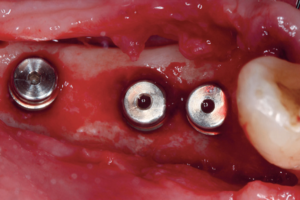
Este caso clínico ilustra a remoção de um implante dentário presente no interior do seio maxilar. Um paciente masculino, de 56 anos de idade, relatou o acidente durante a instalação de um implante dentário na região 26. Localmente, havia uma comunicação bucosinusal, e uma imagem por tomografia computadorizada de feixe cônico (TCFC) revelou o posicionamento do implante. Após os exames e medicamentos pré-operatórios, e uma radiografia panorâmica de auxílio, a técnica de Caldwell-Luc modificada foi usada, com osteotomia na parede lateral do seio maxilar, sendo necessária uma nova TCFC em função do deslocamento desse corpo estranho. Em seguida, a membrana de Schneider foi reparada com membranas L-PRF (fibrina rica em plaquetas e leucócitos), além do levantamento do seio maxilar (aumento de volume ósseo por técnica atraumática do seio maxilar – Tatum) em sessão única, sendo utilizada uma hidroxiapatita como material de enxerto, misturada ao I-PRF. O acesso foi selado com novas camadas de L-PRF. Após a cicatrização (seis meses), um novo implante dentário foi planejado virtualmente e posicionado por meio de cirurgia guiada. Com as etapas restauradoras provisórias concluídas, uma coroa metalocerâmica definitiva foi instalada quatro meses depois. Após 1 ano, o paciente encontra-se sem intercorrências inflamatórias nos tecidos moles e sem mobilidade do implante.
Palavras-chave: enxerto ósseo, complicações intraoperatórias, seio maxilar, remoção de implante, técnica de Caldwell- Luc modificada.
Removal of intrasinusal dental implant, membrane repair and bone grafting at the same surgical session: one year of follow-up
ABSTRACT
This clinical case illustrates the removal of a dental implant from inside the maxillary sinus. A 56-year-old male patient reported the accident during an implant placement procedure in the region of 26. Locally, a bucco-sinusal communication was detected, and a cone beam computed tomography (CBCT) scan revealed its positioning. After systemic evaluation, pre-operative medications, and an auxiliary panoramic radiograph to aid in locating the foreign body, the modified Caldwell-Luc surgical technique was employed, with an osteotomy at the lateral wall of the maxillary sinus. A new CBCT was also required due to the displacement of the dental implant. Next, the Schneiderian membrane was repaired with L-PRF membranes (leukocyte- and platelet-rich fibrin), and a maxillary sinus lift procedure (Tatum bone volume augmentation technique) was performed in a single session, using synthetic hydroxyapatite mixed with liquid injectable i-PRF. The surgical access was sealed with new L-PRF layers. After six months of uneventful healing, a new dental implant was virtually planned and placed using guided surgery. After completing the provisional restorative phases, a definitive metalloceramic crown was delivered four months later. One year later, the patient remains free of soft tissue inflammation, with no mobility at the implant site.
Keywords: intraoperative complications, bone graft, maxillary sinus, implant removal, modified Caldwell-Luc technique.
Referências
1. Manor Y, Anavi Y, Gershonovitch R, Lorean A, Mijiritsky E. Complications and management of implants migrated into the maxillary sinus. Int J Periodontics Restorative Dent. 2018 Nov/Dec;38(6):e112–e118.
2. Garcia CF, Alves RC, Gomes FV, Mayer L. Intercorrência com implantes em seio maxilar relato de caso. Rev Odontol Bras Central. 2017; 26(79): 77-81.
3. An JH, Park SH, Han JJ, Jung S, Kook MS, Park HJ et al. Treatment of dental implant displacement into the maxillary sinus. Maxillofac Plast Reconstr Surg. 2017 Nov 25;39(1):35.
4. Biglioli F, Chiapasco M. An easy access to retrieve dental implants displaced into the maxillary sinus: the bony window technique. Clin Oral Implants Res. 2014 Dec;25(12):1344-51. doi: 10.1111/clr.12276.
5. Bruniera JF, Silva-Sousa YT, Faria PE. Atypical case of three dental implants displaced into the maxillary sinus. Case Rep Dent. 2015;2015:896423.
6. Cavezzi Júnior O. Migration of a dental implant into the maxillary sinus: a case report. Oral Sci. 2015 7(1)3-6.
7. Jeong KI, Kim SG, Oh JS, You JS. Implants displaced into the maxillary sinus: a systematic review. Implant Dent. 2016 Aug; 25(4):547-51.
8. Lee KC, Lee SJ. Clinical features and treatments of odontogenic sinusitis. Yonsei Med J. 2010 Nov;51(6):932-7.
9. Chen YW, Huang CC, Chang PH, Chen CW, Wu CC, Fu CH et al. The characteristics and new treatment paradigm of dental implant-related chronic rhinosinusitis. Am J Rhinol Allergy. 2013 May-Jun;27(3):237-44.
10. Park WB, Han JY, Oh SL. Maxillary sinusitis associated with peri-implantitis at sinus floor augmented sites: case series. Implant Dent. 2019 Oct;28(5):484-489.
11. Manfredi M, Fabbri C, Gessaroli M, Morolli F, Stacchini M. Surgical fenestrated approach to the maxillary sinus like alternative to Caldwell-Luc technique. Minerva Stomatol. 2019 Dec;68(6):308-316.
12. Massuda CKM, Carvalho MR, Miziara LNB, Paiva RS, Marão HF, Pimentel AC et al. Management of perforation of Schneider’s membrane in maxillary sinus lift with L-PRF – case report. RSD [Internet]. 2021Aug.16 [cited 2025May20];10(10):e472101019180.