AUTORES
Renata Faria
Mestra e doutora em Prótese Dentária – Unesp, São José dos Campos; Professora titular do Depto. de Prótese – Unip; Coordenadora da área de Prótese do curso de especialização em Implantodontia – Unimes.
Orcid: 0000-0001-6770-2734.
Joyce Roma Correia dos Santos Siqueira
Especialista em Implantodontia – APCD; Especialista em Prótese Dentária – Uningá; Mestranda no programa de pós-graduação de Ciências Aplicadas à Saúde Bucal – Unesp, São José dos Campos.
Orcid: 0000-0002-6733-1643.
Rita Maria Morejon Rodriguez
Graduada em Odontologia – UCMM, Cuba; Mestranda no programa de pós-graduação de Ciências Aplicadas à Saúde Bucal – Unesp, São José dos Campos.
Orcid: 0000-0002-4218-1079.
RESUMO
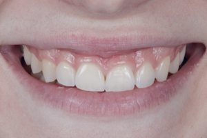
O fluxo digital permite ao profissional proporcionar maior conforto, agilidade, precisão e menor número de visitas do paciente ao consultório, já que com o uso de ferramentas digitais, como o scanner e as impressoras 3D, o processo de confecção de modelos provisórios e placas é feito em menor tempo e com maior previsibilidade. Desta forma, grandes e complexas reabilitações, como no caso de pacientes com diminuição da dimensão vertical devido a diversos fatores, como desgaste químico ou mecânico, ou ainda ausências dentárias, são possíveis de serem realizadas de maneira eficiente, rápida e precisa. Desta maneira, o objetivo do presente artigo foi a apresentação de um caso clínico através do planejamento e execução de uma reabilitação total dos arcos superior e inferior com restaurações cerâmicas, utilizando o fluxo digital como ferramenta para a confecção desde o mock-up até a placa estabilizadora.
Palavras-chave – IOS; CAD/CAM; Scanner intraoral; Odontologia digital; Reabilitação bucal.
ABSTRACT
The digital workflow allows the professional to provide greater comfort, agility, precision and fewer visits by the patient to the office, since with the use of digital tools such as the scanner and 3D printers, the process of making models, provisionals and occlusal devices is done in less time and with greater predictability. In this way, large and complex rehabilitations, as in the case of patients with reduced vertical dimension due to several factors such as wear, whether chemical or mechanical, or even missing teeth, are possible to be performed efficiently, quickly and accurately. Thus, the objective of this article is to present a clinical case through the planning and execution of a total rehabilitation of the upper and lower arches with ceramic restorations, using the digital worflow as a tool for making from the mock up to the stabilizing occlusal device.
Key words – IOS; CAD/CAM; Intraoral scanner; Digital dentistry; Oral rehabilitation.
Recebido em mar/2022
Aprovado em mar/2022
Referências
- Johansson A, Johansson AK, Omar R, Carlsson GE. Rehabilitation of the worn dentition. J Oral Rehabil 2008;35(7):548-66.
- Shellis RP, Addy M. The interactions between attrition, abrasion and erosion in tooth wear. Monogr Oral Sci 2014;25:32-45 (DOI: 10.1159/000359936).
- De Boever JA, Carlsson GE, Klineberg IJ. Need for occlusal therapy and prosthodontic treatment in the management of temporomandibular disorders. Part II. Tooth loss and prosthodontic treatment. J Oral Rehabil 2000;27(8):647-59.
- Oliveira GAS, Beatrice LCS, Leão SFS. Reabilitação oral em pacientes com bruxismo: o papel da Odontologia Restauradora. Int J Dent 2007;6(4):117-23.
- Olthoff LW, Van Der Glas HW, Van Der Bilt A. Influence of occlusal vertical dimension on the masticatory performance during chewing with maxillary splints. J Oral Rehabil 2007;34(8):560-5.
- Blatz MB, Conejo J. The current state of chairside digital dentistry and materials. Dent Clin North Am 2019;63(2):175-97.
- Papadopoulos C, Dionysopoulos D, Tolidis K, Kouros P, Koliniotou-Koumpia E, Tsitrou E. Structural integrity evaluation of large MOD restorations fabricated with a bulk-fill and a CAD/CAM resin composite material. Oper Dent 2019;44(3):312-21.
- Kömürcüoğlu MB, Sağırkaya E, Tulga A. Influence of different surface treatments on bond strength of novel CAD/CAM restorative materials to resin cement. J Adv Prosthodont 2017;9(6):439-46.
- Rivera-Morales WC, Mohl ND. Restoration of the vertical dimension of occlusion in the severely worn dentition. Dent Clin North Am 1992;36(3):651-64.
- Dawson PE. New definition for relating occlusion to varying conditions of the temporomandibular joint. J Prosthet Dent 1995;74(6):619-27.
- Baheti MJ, Soni UN, Gharat NV, Mahagaonkar P, Khokhani R, Dash S. Intra-oral scanners: a new eye in dentistry. Austin J Orthopade & Rheumatol 2015;2(3):1021.
- Di Fiore A, Meneghello R, Graiff L, Savio G, Vigolo P, Monaco C et al. Full arch digital scanning systems performances for implant-supported fixed dental prostheses: a comparative study of 8 intraoral scanners. J Prosthodont Res 2019;63(4):396-403.
- Winkler J, Gkantidis N. Trueness and precision of intraoral scanners in the maxillary dental arch: an in vivo analysis. Sci Rep 2020;10(1):1172.
- Tomita Y, Uechi J, Konno M, Sasamoto S, Iijima M, Mizoguchi I. Accuracy of digital models generated by conventional impression/plaster-model methods and intraoral scanning. Dent Mater J 2018;37(4):628-33.
- Rapone B, Palmisano C, Ferrara E, di Venere D, Albanese G, Corsalini M. The accuracy of three intraoral scanners in the oral environment with and without saliva: a comparative study. Appl Sci 2020;10(21):7762.
- Chen Y, Zhai Z, Li H, Yamada S, Matsuoka T, Ono S et al. Influence of liquid on the tooth surface on the accuracy of intraoral scanners: an in vitro study. J Prosthodont 2022;31(1):59-64.
- Vág J, Renne W, Revell G, Ludlow M, Mennito A, Teich ST et al. The effect of software updates on the trueness and precision of intraoral scanners. Quintessence Int 2021;52(7):636-44.
- Rivera-Morales WC, Mohl ND. Relationship of occlusal vertical dimension to the health of the masticatory system. J Prosthet Dent 1991;65(4):547-53.
- Ferrario VF, Sforza C, Serrao G, Schmitz JH. Three-dimensional assessment of the reliability of a postural face-bow transfer. J Prosthet Dent 2002;87(2):210-5.
- LeSage BP. CAD/CAM: applications for transitional bonding to restore occlusal vertical dimension. J Esthet Restor Dent 2020;32(2):132-40.
- Nanda A, Jain V, Manak K, Verma M. An alternative adhesive based technique of raising the occlusal vertical dimension. Indian J Dent Res 2014;25(4):505-8.
- Nanda A, Jain V, Srivastava A. An electromyographic study to assess the minimal time duration for using the splint to raise the vertical dimension in patients with generalized attrition of teeth. Indian J Dent Res 2011;22(2):303-8.
- Zhang Y, Kelly JR. Dental ceramics for restoration and metal veneering. Dent Clin North Am 2017;61(4):797-819.
- Cagidiaco EF, Grandini S, Goracci C, Joda T. A pilot trial on lithium disilicate partial crowns using a novel prosthodontic functional index for teeth (FIT). BMC Oral Health 2019;19(1):276.
- Gehrt M, Wolfart S, Rafai N, Reich S, Edelhoff D. Clinical results of lithium-disilicate crowns after up to 9 years of service. Clin Oral Investig 2013;17(1):275-84.
- Pieger S, Salman A, Bidra AS. Clinical outcomes of lithium disilicate single crowns and partial fixed dental prostheses: a systematic review. J Prosthet Dent 2014;112(1):22-30.
- Ma L, Guess PC, Zhang Y. Load-bearing properties of minimal-invasive mono- lithic lithium disilicate and zirconia occlusal onlays: finite element and theoretical analyses. Dent Mater 2013;29(7):742-51.
- Okeson JP. Orofacial pain: guidelines for assessment, diagnosis and management. Carol Stream (IL): Quintessence, 1996. p.1-14.
- Greene CS, Laskin DM. Splint therapy for the myofascial pain-dysfunction (MPD) syndrome: a comparative study. J Am Dent Assoc 1972;84(3):624-8.
- Carraro JJ, Caffesse RG. Effects of occlusal splints on TMJ symptomatology. J Prosthet Dent 1978;40(5):563-6.