Trabalho relata um caso clínico de maxila apresentava-se severamente atrófica e com pneumatização dos seios maxilares bilateralmente.
AUTORES
Rafael Zetehaku Araújo
Doutorado em Implantodontia e Prótese sobre Implante – Universidade Federal de Uberlândia; Mestre em Cirurgia Bucomaxilofacial – Universidade do Sagrado Coração; Especialista em Cirurgia Bucomaxilofacial – Hospital das Clínicas da Universidade Federal de Minas Gerais.
Orcid: 0000-0002-2893-0408.
Rosenvaldo Moreira Júnior
Especialização em Implantodontia – Factese; Doutor em Cirurgia Bucomaxilofacial – Universidade do Sagrado Coração.
Orcid: 0000-0002-2210-3006.
João Vitor Lemos Pinheiro
Aperfeiçoamento em Cirurgia e Traumatologia Bucomaxilofacial, e Implantodontia – Núcleo de Ensino e Estética em Odontologia; Graduado em Odontologia – Faculdade de Ilhéus.
Orcid: 0000-0002-0051-471X.
João Carlos Leahy
Especialista em Implantodontia – Universidade Federal da Bahia; Mestrando em Implantodontia – São Leopoldo Mandic.
Orcid: 0000-0003-3596-0768.
Marcos Martins Curi
Mestre e doutor em Patologia – Fundação Antônio Prudente; Especialista em Cirurgia Bucomaxilofacial – Hospital do Câncer de São Paulo; Coordenador do serviço de Cirurgia Bucomaxilofacial – Hospital Santa Catarina.
Orcid: 0000-0001-8216-3564.
RESUMO
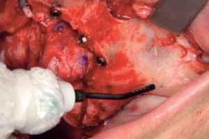
O objetivo deste trabalho foi relatar um caso clínico. Uma paciente do sexo feminino, com 54 anos de idade, compareceu ao ambulatório da pós-graduação em Implantodontia do Núcleo de Ensino e Estética em Odontologia (Itabuna/BA, Brasil) com o objetivo de “recuperar a arcada dentária”. No exame clínico e radiográfico, a maxila apresentava-se severamente atrófica e com pneumatização dos seios maxilares bilateralmente. O tratamento proposto foi a reabilitação com cirurgia guiada e uma combinação de ancoragens esqueléticas em pilar canino com instalação de implantes angulados e “approach palatino”, mas, principalmente, a utilização dos pilares pterigoides para ancoragem posterior, permitindo a reabilitação protética através de carga imediata. O planejamento virtual foi realizado planejando-se a instalação de implantes na região anterior de maxila e dois implantes pterigoides bilateralmente. Realizada a cirurgia e com o travamento de todos os implantes acima de 40 Ncm, a confecção da prótese foi realizada em carga imediata com ajustes oclusais progressivos, proporcionando estabilidade funcional e estética.
Palavras-chave – Maxila atrófica; Implante pterigoide; Cirurgia guiada.
ABSTRACT
The objective of this work is to report a clinical case, whose patient, female, 54 years old, attended the post-graduate outpatient clinic in Implantology at the Center for Teaching and Aesthetics in Dentistry (Itabuna/BA, Brazil) with the aim of “recovering the dental arch”. On clinical and radiographic examination, the maxilla was severely atrophic. The proposed treatment was rehabilitation with guided surgery with a combination of skeletal anchorages in a canine abutment with the installation of angled implants and a “palatal approach”, but mainly, the use of pterygoid abutments for posterior anchoring, allowing prosthetic rehabilitation through “immediate loading”. The virtual planning consisted on implant placement in the anterior region of the maxilla and two pterygoid implants bilaterally. After the surgery and with final insertion torques above 40 Ncm, the prosthesis was delivered under immediate loading, with progressive occlusal adjustments, providing functional and esthetic stability.
Key words – Atrophic maxilla; Pterygoid implant; Guided surgery.
Recebido em out/2020
Aprovado em nov/2021
Referências
- Adell R, Lekholm U, Rockler B, Brånemark P-I. A 15-year study of osseointegrated implants in the treatment of the edentulous jaw. Int J Oral Surg 1981;10(6):387-416.
- Balshi SF, Wolfingen GJ, Balshi TJ. Prosthodontic revision treatment: total oral rehabilitation of a patient with intense parafunction and advanced bone loss in the posterior maxilla. ImplantNews 2014;11(6a):24-39.
- Curi MM, Cardoso CL, Ribeiro KCB. Retrospective study of pterygoid implants in the atrophic posterior maxilla: implant and prosthesis survival rates up to 3 years. Int J Oral Maxillofac Implants 2015;30(2):378-83.
- Gomez-Polo M, Ortega R, Gomez-Polo C, Martin C, Celemin A, Rio JD. Does length, diameter, or bone quality affect primary and secondary stability in self-tapping dental implants? J Oral Maxillofac Surg 2016;74(7):1344-53.
- Lekholm U, Zarb G. Patient selection and preparation. In: Brånemark PI, Zarb G, Albrektsson T. Editors. Tissue-integrated prostheses. Chicago: Quintessence, 1985. p.199-209.
- Albrektsson T, Zarb GA, Worthington P, Eriksson A. The long-term efficacy of currently used dental implants: a review and proposed criteria of success. Int J Oral Maxillofac Implants 1986;1(1):11-26.
- Chiapasco M. Early and immediate restoration and loading of implants in completely edentulous patients. Int J Oral Maxillofac Implants 2004;19(suppl.):76-91.
- Goiato MC, dos Santos DM, Santiago Jr. JF, Moreno A, Pellizzer EP. Longevity of dental implants in type IV bone: a systematic review. Int J Oral Maxillofac Surg 2014;43(9):1108-16.
- Adell R, Eriksson B, Lekholm U, Brånemark P-I, Jemt T. A long-term follow-up study of osseointegrated implants in the treatment of totally edentulous jaws. Int J Oral Maxillofac Implants 1990;5(4):347-59.
- Balshi SF, Wolfinger GJ, Balshi TJ. Analysis of 164 titanium oxide-surface implants in completely edentulous arches for fixed prosthesis anchorage using the pterygomaxillary region. Int J Oral Maxillofac Implants 2005;20(6):946-52.
- Araujo RZ, Santiago-Júnior JF, Cardoso CL, Condezzo AFB, Moreira-Júnior R, Curi MM. Clinical outcomes of pterygoid implants: systematic review and meta-analysis. J Cranio Maxillofac Surg 2019;47(4):651-
- Langer B, Langer L, Herrman I, Jorneous L. The wide fixture: a solution for special bone situations and a rescue for the compromised implant. Int J Oral Maxillofac Implants 1993;8(4):400-08.
- Ridell A, Grondahl K, Sennerby L. Placement of Brånemark implants in the maxillary tuber region: anatomical considerations, surgical technique and long-term results. Clin Oral Implants Res 2009;20(1):94-8.
- Tulasne JF. Osseointegrated fixtures in the pterygoid region. In: Worthington P, Brånemark PI (eds). Advanced Osseointegration Surgery. Applications in the Maxillofacial Region. Chicago: Quintessence, 1992. p.182-8.
- Balshi SF, Wolfinger GJ, Balshi TJ. Surgical planning and prosthesis construction using computer technology and medical imaging for immediate loading of implants in the pterygomaxillary region. Int J Periodontics Restorative Dent 2006;26(3):239-47.
- Valerón JF, Valerón PF. Long-term results in placement of screw-type implants in the pterygomaxillary-pyramidal region. Int J Oral Maxillofac Implants 2007;22(2):195-200.
- Rodriguez X, Mendez V, Vela X, Segala M. Modified surgical protocol for placing implants in the pterygomaxillary region: clinical and radiologic study of 454 implants. Int J Oral Maxillofac Implants 2012;27(6):1547-53.
- Balshi TJ, Wolfinger GJ, Slauch RW, Balshi SF. A retrospective comparison of implants in the ptergygomaxillary region: implant placement with two-stage, single-stage, and guided surgery protocols. Int J Oral Maxillofac Implants 2013;28(1):184-
- World Health Organization. International classification of functioning, disability and health: ICF. Geneva: World Health Organization, 2001.
- Lee SP, Paik KS, Kim MK. Anatomical study of the pyramidal process of the palatine bone in relation to implant placement in the posterior maxilla. J Oral Rehabil 2001;28(2):125-32.
- Rodriguez X, Lucas-Taulé E, Elnayef B, Altuna P, Gargallo-Albiol J, Penarrocha MD et al. Anatomical and radiological approach to pterygoid implants: a cross-sectional study of 202 cone beam computed tomography examinations. Int J Oral Maxillofac Surg 2016;45(5):636-40.
- Rodriguez X, Rambla F, De Marcos Lopez L, Mendez V, Vela X, Jimenez Garcia J. Anatomical study of the pterygomaxillary area for implant placement: cone beam computed tomographic scanning in 100 patients. Int J Oral Maxillofac Implants 2014;29(5):1049-52.
- Balshi TJ, Lee Y, Hernandez RE. The use of pterygoid implants in the partially edentulous patient: a preliminary report. Int J Oral Maxillofac Surg 1995;10(1):89-98.
- Bidra AS, Huynh-Ba G. Implants in the pterygoid region: a systematic review of the literature. Int J Oral Maxillofac Surg 2011;40(8):773-81.
- Candel E, Peñarrocha D, Peñarrocha M. Rehabilitation of the atrophic posterior maxilla with pterygoid implants: a review. J Oral Implantol 2012;38(spec):461-6.
- Krekmanov L. Placement of posterior mandibular and maxillary implants in patients with severe bone deficiency: a clinical report of procedure. Int J Oral Maxillofac Implants 2000;15(5):722-30.
- Barbeiro RH, Medeiros FRM, Buzza EPN, Magda A, Coppedê AR, Shibli JA. Tratamento de maxilas atróficas por meio de fixações zigomáticas sob carregamento imediato: relatos de coorte histórico com 8-60 meses de acompanhamento. ImplantNews 2015;12(5):589-96.
- Duarte LR, Peredo LG, Nary Filho H, Francischone CE, Brånemark PI. Reabilitação da maxila atrófica utilizando quatro fixações zigomáticas em sistema de carga imediata. ImplantNews 2004;1(1):45-50.
- Graves SL. The pterygoid plate implant: a solution for restoring the posterior maxilla. Int J Periodontics Restorative Dent 1994;14(6):512-23.
- Peñarrocha M, Carrillo C, Boronat A, Peñarrocha M. Retrospective study of 68 implants placed in the pterygomaxillary region using drills and osteotomes. Int J Oral Maxillofac Implants 2009;24(4):720-6.