Artigo apresenta técnica minimamente invasiva para a coleta do periósteo do palato, possibilitando o isolamento e a expansão das células-tronco mesenquimais.
AUTORES
José R. M. Ferreira
Cirurgião-dentista – Ufes; Especialista em Periodontia – PUC-Campinas; Mestre em Implantodontia – Unigranrio; Doutor em Ciências dos Materiais pelo IME; Fundador, pesquisador e presidente do Centro de Processamento Celular R-Crio Criogenia S.A.
Orcid: 0000-0002-5192-5623.
Tamara C. L. Castro
Farmacêutica – USF; Depto. de Bioengenharia – R-Crio Criogenia S.A.
Orcid: 0000-0002-5018-4405.
Anna Carolina M. Ferreira de Almeida
Médica veterinária – UniFAJ; Depto. de Bioengenharia – R-Crio Criogenia S.A.
Orcid: 0000-0002-4391-4211.
Daniel N. da Rocha
Cirurgião-dentista – Estácio de Sá; Mestre e doutor em Ciências dos Materiais – IME; Depto. de Bioengenharia – R-Crio Criogenia S.A.
Orcid: 0000-0003-2413-345X.
André A. Pelegrine
Cirurgião-dentista – PUC-Campinas; Especialista em Periodontia e professor – São Leopoldo Mandic; Mestre em Implantodontia – Unisa; Doutor em Clínica Médica – Unicamp; Pós-doutorado – Unifesp.
Orcid: 0000-0001-7935-1062.
RESUMO
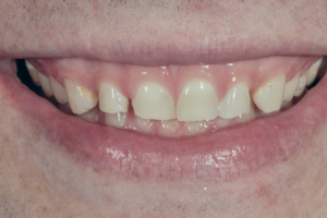
Neste trabalho, uma técnica minimamente invasiva para a coleta do periósteo do palato é apresentada, possibilitando o isolamento e a expansão das células-tronco mesenquimais em qualquer fase da vida do indivíduo. O doador com 77 anos de idade foi submetido à biopsia do tecido do palato duro por meio de um bisturi circular, com 3 mm de diâmetro, nas proximidades da região dos pré-molares superiores, sem a necessidade da realização de suturas, devido ao tamanho mínimo de incisão. A cultura celular do periósteo proveniente do palato foi estabelecida com sucesso, chegando ao final da quarta passagem após 53 dias de cultivo. A análise por citometria de fluxo indicou que o periósteo do palato contém população de células-tronco mesenquimais CD73, CD90 e CD105 positivas, além da baixa expressão de marcadores de superfície de células-tronco hematopoiéticas. A expressão do marcador CD105 (76,10%) indica potencial osteogênico dessa fonte de células-tronco mesenquimais, o que foi consubstanciado após indução à diferenciação osteogênica. O periósteo do palato demonstrou-se uma fonte satisfatória de células-tronco mesenquimais de fenótipo “jovem”, mesmo em adultos com idade avançada, e com potencial de aplicabilidade na bioengenharia óssea.
Palavras-chave – Células-tronco; Periósteo; Técnicas de cultura de células.
ABSTRACT
In this work, a minimally invasive technique for collecting the periosteum from the palate is presented, enabling the isolation and expansion of mesenchymal stem cells at any stage of the individual’s life. The 77-year-old donor underwent hard palate tissue biopsy using a circular scalpel, 3 mm in diameter, in the vicinity of the upper premolar region, without the need to perform sutures, due to the minimal size incision. Cell culture of periosteum from the palate was successfully established, reaching the end of the 4th passage after 53 days of culture. Flow cytometric analysis indicated that the palate periosteum contains a population of CD73, CD90 and CD105 positive mesenchymal stem cells, in addition to the low expression of hematopoietic stem cell surface markers. The expression of the CD105 marker (76.10%) indicates the osteogenic potential of this source of mesenchymal stem cells, which was substantiated after induction of osteogenic differentiation. The palate periosteum proved to be a satisfactory source of mesenchymal stem cells of the “young” phenotype, even in adults with advanced age, and with potential applicability in bone bioengineering.
Key words – Stem cells; Periosteum; Cell culture techniques.
Recebido em nov/2021
Aprovado em dez/2021
Referências
- Oliver JD, Madhoun W, Graham EM, Hendrycks R, Renouard M, Hu MS. Stem cells regenerating the craniofacial skeleton: current state-of-the-art and future directions. J Clin Med 2020;9(10):3307.
- Gronthos S, Mankani M, Brahim J, Robey PG, Shi S. Postnatal human dental pulp stem cells (DPSCs) in vitro and in vivo. Proc Natl Acad Sci USA 2000;97(25):13625-30.
- Schmelzeisen R, Schimming R, Sittinger M. Making bone: implant insertion into tissue-engineered bone for maxillary sinus floor augmentation – a preliminary report. J Craniomaxillofac Surg 2003;31(1):34-9.
- Egusa H, Sonoyama W, Nishimura M, Atsuta I, Akiyama K. Stem cells in dentistry – part I: stem cell sources. J Prosthodont Res 2012;56(3):151-65.
- Ferretti C, Mattioli-Belmonte M. Periosteum derived stem cells for regenerative medicine proposals: boosting current knowledge. World J Stem Cells 2014;6(3):266-77.
- Rocha DN, Rlsb M, Barbosa RM. Mesenchymal stem cells associated with bioceramics for bone tissue regeneration. Biomater Med Appl 2017;1(2):1-8.
- Mitsiadis TA, Feki A, Papaccio G, Catón J. Dental pulp stem cells, niches, and notch signaling in tooth injury. Adv Dent Res 2011;23(3):275-9.
- Al-Qtaitat A, Shore RC, Aaron JE. Structural changes in the ageing periosteum using collagen III immuno-staining and chromium labelling as indicators. J Musculoskelet Neuronal Interact 2010;10(1):112-23.
- Nahian A, Chauhan PR. Histology, periosteum and endosteum. Treasure Island: StatPearls Publishing, 2022.
- Block TJ, Marinkovic M, Tran ON, Gonzalez AO, Marshall A, Dean DD et al. Restoring the quantity and quality of elderly human mesenchymal stem cells for autologous cell-based therapies. Stem Cell Res Ther 2017;8(1):239.
- Yukata K, Xie C, Li TF, Takahata M, Hoak D, Kondabolu S et al. Aging periosteal progenitor cells have reduced regenerative responsiveness to bone injury and to the anabolic actions of PTH 1-34 treatment. Bone 2014;62(1):79-89.
- Iezzi I, Lazzarini R, Cerqueni G, Hosein A, Rossato M, Licini C et al. MicroRNA profiling in mesenchymal stromal cells: the tissue source as the missing piece in the puzzle of ageing. Stem Cell Rev Rep 2021;17(3)1014-26.
- Naung NY, Duncan W, Silva RD, Coates D. Localization and characterization of human palatal periosteum stem cells in serum‐free, xeno‐free medium for clinical use. European J Oral Sci 2019;127(2):99-111.
- Ishiy FAA, Fanganiello RD, Kobayashi GS, Kague E, Kuriki PS, Passos-Bueno MR. CD105 is regulated by hsa-miR-1287 and its expression is inversely correlated with osteopotential in SHED. Bone 2018;106:112-20.
- Naung NY, Duncan WJ, De Silva RK, Coates DE. HGF/MET in osteogenic differentiation of primary human palatal periosteum-derived mesenchymal stem cells. J Oral Sci 2021;63(4):341-6.