AUTORES
Karla Valéria Marques Rocha Lima
Mestranda em Implantodontia – Faculdade São Leopoldo Mandic; Cirurgiã-dentista especialista em Implantodontia.
Orcid: 0000-0003-1281-4768.
Roberto Brunow Lehmann
Professor do Depto. de Engenharia Metalúrgica – Faculdade de Engenharia da Universidade Federal Fluminense.
Orcid: 0000-0001-9319-3179.
Liliane Pacheco de Carvalho
Professora do Depto. de Odontologia – Centro Universitário Governador Ozanam Coelho e Faculdade de Odontologia da São Leopoldo Mandic.
Orcid: 0000-0002-3837-7618.
Ana Elisa de Oliveira Matos
Professora do Depto. de Odontologia Restauradora – Universidade Federal de Juiz de Fora.
Orcid: 0000-0002-7116-2336.
Bruno Salles Sotto Maior
Professor do Depto. de Odontologia Restauradora – Universidade Federal de Juiz de Fora e Faculdade de Odontologia da São Leopoldo Mandic.
Orcid: 0000-0002-9462-0299.
RESUMO
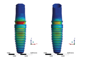
Objetivo: comparar biomecanicamente as diferentes alturas dos componentes protéticos em implantes cone-morse extraestreitos na região do incisivo lateral superior, quando submetidos a forças oblíquas. Material e métodos: foram criados quatro modelos com implantes de diâmetro 2,9 mm x 11,5 m de comprimento na região do incisivo lateral superior com próteses de pilares de 3,3 mm de diâmetro, com altura de cimentação de 6 mm e alturas de transmucoso de 0,8 mm, 1,5 mm, 3,5 mm e 4,5 mm, através do método de elementos finitos utilizando o software Ansys Workbench versão 14. Para o carregamento sobre a prótese, foi utilizado um aplicador de carga localizado a 3 mm da borda incisal da face palatina, com intensidade de força de 100 N e 30o de angulação. Resultados: houve diferenças significativas de tensão para os quatro pilares simulados, tanto na prótese como no implante e no osso. Contudo, os maiores valores obtidos não sugerem falha no sistema por ficarem abaixo do limite de escoamento dos materiais. Conclusão: apesar do aumento de tensões nos pilares mais altos no conjunto pilar/ implante/osso, todas as alturas suportam as tensões e não sugerem falha no sistema.
Palavras-chave – Implantes extraestreitos; Plataforma cone- -morse; Elementos finitos; Análise de tensões.
ABSTRACT
Objective: to biomechanically compare the different heights of prosthetic abutments for extra-narrow Morse taper dental implants in the maxillary lateral incisor region, when subjected to oblique forces. Material and methods: four models were created with (2.9 mm x 11.5 m in length) implants in the lateral incisor region. The prosthetic abutments were 3.3 mm in diameter and 6 mm in height, presenting transmucosal collars of 0.8 mm, 1.5 mm, 3.5 mm, and 4.5 mm. All prostheses were loaded 3 mm from the incisal edge of the palatal aspect under 100 N and 30 degrees of angulation using the finite element methods (Ansys Workbench, software version 14). Results: there were significant differences in tension values among all four simulated abutments (prostheses, dental implant, and the bone components). However, the highest values generated did not suggest system failure because they remained below the yield point of all tested materials. Conclusion: despite the increase in tensions in the longest abutments at the abutment/implant/bone set, all heights supported the stresses and did not suggest system failure.
Key words – Extra narrow implants; Cone morse connection; Finite element analyis; Stress analysis.
Recebido em nov/2021
Aprovado em fev/2022
Referências
- Slagter KW, Hartog L, Bakker NA, Vissink A, Meijer HJ, Raghoebar GM. Immediate placemment of dental implants in the esthetic zone: a systematic review and pooled analysis. J Periodontol 2014;85(7):241-50.
- Tarnow DP, Cho SC, Wallace SS. The effect of inter-implant distance on the height of inter-implant bone crest. J Periodontol 2000;71(4):546-9.
- Rosa AC, Rosa JC, Pereira LAD, Francischone CE, Sottomaior BS. Guidelines for selecting the implant diameter during immediate implant placement of a fresh extraction socket: a case series. Int J Periodontics Restorative Dent 2016;36(3):401-7.
- Capelli M, Testori T, Galli F, Motroni A, Weisnstein R, Zuffetti F et al. Implant-buccal plate distance as diagnostic parameter: a prospective cohort study on implant placement in fresh extraction sockets. J Periodontol 2013;84(12):1768-74.
- Kang JS, Heo HY, Son MK. Preliminary study for fully digital guided implant treatment with narrow implants in the anterior region. Oral Biology Research 2020;44(3):109-17.
- Schiegnitz E, Al-Nawas B. Narrow-diameter implants: a systematic review and meta-analysis. Clin Oral Implants Res 2018;29(suppl.16):21-40.
- Gonzalez-Valls G, Rocha-Millan E, Cespedes-Sanches JM, Gonzalez-Navarro B, Torrejon-Moya A, Lopez-Popez J. Narrow diameter dental implants as an alternative treatment for atrophic aveolar ridges. Systematic review and meta-analysis. Materials (Basel) 2021;14(12):3234.
- Al-Johany SS, Al Amri MD, Alsaeed S, Alalola B. Dental implant lenght and diameter. A proposed classification scheme. J Prosthodont 2017;26(3):252-60.
- Lee BA, Kim BH, Kweon HHI, Kim YT. The prosthetic abutment height can affect marginal bone loss around dental implants. Clin Implant Dent Relat Res 2018;20(5):799-805.
- Galindo-Moreno P, Leon-Cano A, Ortega-Oller I, Monje A, Suarez F, Ovalle F et al. Prosthetic abutment height is a key fator in peri-implant marginal bone loss. J Dent Res 2014;93(7):80-5.
- Geng JP, Tan KB, Liu GR. Application of finite element analysis in implant dentistry: a review of the literature. J Prostet Dent 2001;85(6):585-98.
- Saab XE, Griggs JA, Powers JM, Engelmeier RL. Effect of abutment angulation on the strain on the bone around na implant in the anterior maxila: a finite element study. J Prosthet Dent 2017;97(2):85-92.
- Çaglar A, Bal BT, Aydin C, Yilmaz H, Ozkan S. Evaluation of stress occurring one three different zirconia dental implants: three dimensional finite element analysis. Int J Oral Maxillofac Implants 2010;25(1):95-103.
- Ferreira EM. Análise biomecânica em prótese fixa posterior de três elementos em implantes com três tipos diferentes de conexão pelo método de elemento finito [tese]. Campinas: São Leopoldo Mandic, 2012.
- Calvário MAF. Análise comparativa da distribuição das tensões em prótese cimentada e parafusada entre implantes unitários retos e inclinados submetidos a diferentes tipos de cargas pelo método de elementos finitos [tese]. Campinas: Centro de Pesquisas Odontológicas São Leopoldo Mandic, 2011.
- Lemos CAA, Verri FR, Bonfante EA, Santiago JF, Pellizzer EP. Comparison of external and internal implant–abutment connections for implant supported prostheses. A systematic review and meta-analysis. J Dent 2018;70:14-22.
- Albuquerque M. Análise comparativa das tensões por elementos finitos entre implantes de diâmetro reduzido regular [tese]. Campinas: São Leopoldo Mandic, 2015.
- Çiftçi Y, Canay S. The effect of veneering materials on stress distribution in implant-supported fixed prosthetic restorations. Int J Oral Maxillofac Implants 2000;15(4):571-82.
- Bourarel C, Aitlahrach M, Heinemann F, Hasan I. Biomechanical finite element analysis of small diameter and short dental implants. Extensive study of commercial implants. Biomed Tech (Berl) 2012;57(1):21-32.
- Akça K, Cehreli MC, Iplikçioğlu H. Evaluation of the mechanical characteristics of the implant-abutment complex of a reduced-diameter morse-taper implant. A nonlinear finite element stress analysis. Clin Oral Implants Res 2003;14(4):444-54.
- Renouard F, Nissand D. Impact of implant lenght and diameter on survival rates. Clin Oral Implants Res 2006;17(2):35-51.
- Almeida CA. Implantes estreitos e suas utilizações na Implantodontia, suas capacidades e restrições [tese]. Ipatinga: Faculdade de Sete Lagoas, 2020.
- Salvi GE, Lang NP. Changing paradigms in implant dentistry. Crit Rev Oral Biol Med 2001;12(3):262-72.
- Adell R, Lekholm U, Rocler B, Brånemark PI. A 15 yaers study of osseointegrated implants in the treatment of endentulous jaw. Int J Oral Surg 1982;10(6):387-416.
- Qian L, Todo M, Matsushita Y, Koyano K. Effects of implant diameter, insertion depth, and loading angle on stress/strain fields in implant/jawbone systems: finite element analysis. Int J Oral Maxillofac Implants 2009;24(5):877-8.
- Toniollo MB, Macedo AP, Rodrigues RC, Ribeiro RF, Mattos MG. Three-dimensional finite element analysis of stress distribution on different bony ridges with different lengths of morse taper implants and prosthesis dimensions. J Craniofac Surg 2012;23(6):1888-92.
- Mangano C, Shibli JA, Pires JT, Luongo G, Piattelli A, Lezzi G. Early bone formation around immediately loaded transitional implants inserted in the human posterior maxilla: the effects of fixture design and surface. Biomed Res Int 2017;2017:4152506.
- Rodrigues ES, Menegon A, Benetti P, Machado KH. Resistência à fratura da conexão morse friccional considerando-se elementos finitos: revisão da literatura. Salusvita 2020;39(2):549-64.
- Bordin D, Bergamo ETP, Fardin VP, Coelho PG, Bonfante EA. Fracture strength and probability of survival of narrow and extra-narrow dental implants after fatigue testing: in vitro and in silico analysis. J. Mech Behav Biomed Mater 2017;71:244-9.