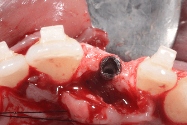
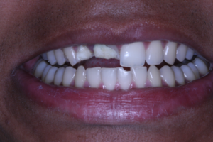
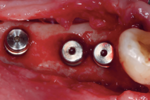
Expansão óssea: artigo demonstra caso clínico de paciente com rebordo severamente reabsorvido na região do 21, com tratamento cirúrgico em sessão única.
AUTORES
Fausto Frizzera
Doutor em Implantodontia, mestre, especialista e pós-doutor em Periodontia – FOAr/Unesp; Professor titular de Periodontia e Implantodontia – Faesa Centro Universitário.
Orcid: 0000-0002-0027-6686.
Mario Groisman
Mestre em Ciências Dentais – Universidade de Lund, Suécia; Especialista em Periodontia – Uerj; Especialista em Implantodontia – CFO.
Orcid: 0000-0003-0202-4156.
Nicolas Nicchio
Graduado em Odontologia – Faesa; Mestrando em Periodontia – FOAr/Unesp.
Orcid: 0000-0001-9332-8862.
Quézia Godinho de Oliveira
Graduada em Odontologia – Ufes; Especialista em Dentística – Uningá.
Orcid: 0000-0001-6148-8968.
Flávia Vilarino Esteves
Graduada em Odontologia – Ufes; Especialista em Prótese Dentária – USP.
Orcid: 0000-0002-5044-2363.
RESUMO
Esse artigo teve como objetivo demonstrar o caso clínico de uma paciente jovem com rebordo severamente reabsorvido na região do 21, cujo tratamento cirúrgico foi realizado em uma sessão única. O procedimento consistiu na expansão do rebordo com instrumentos rotatórios, instalação de implante e regeneração óssea com biomateriais que foram devidamente estabilizados com tachinhas, para permitir maior previsibilidade na recuperação do tecido previamente perdido. Após o período de cicatrização, observou-se a necessidade de realizar uma gengivectomia na região do 23 e de enxertia de tecido mole na região do 21 para se obter um resultado mais estético. O condicionamento tecidual foi dado pelas coroas provisórias e, em seguida, foi realizado um escaneamento digital para confecção de um pilar de zircônia e cimentação de uma coroa de dissilicato de lítio.
Palavras-chave – Implante dentário; Regeneração óssea; Gengiva; Estética; Biomateriais.
ABSTRACT
This article aims to demonstrate a clinical case of a young patient with severely resorbed ridge in the region of 21, where the surgical treatment was performed in a single session. The procedure consisted of the expansion of the ridge with rotating instruments, implant installation and bone regeneration with biomaterials that were properly stabilized with titanium tacks to allow greater predictability in the recovery of previously lost tissue. After the healing period, it was observed the need to perform a gingivectomy in the region of 23 and soft tissue grafting in the region of 21 in order to obtain a more aesthetic result. Tissue conditioning was performed by the temporary crowns and then a digital scan was performed to make a zirconia abutment and cementation of a lithium disilicate crown.
Key words – Dental implant; Bone regeneration; Gingiva; Esthetics; Biomaterials.
Recebido em ago/2021
Aprovado em ago/2021
Referências
- Albrektsson T, Zarb G, Worthington P, Eriksson AR. The long-term efficacy of currently used dental implants: a review and proposed criteria of success. Int J Oral Maxillofac Implants 1986;1(1):11-25.
- Slagter KW, Meijer HJA, Bakker NA, Vissink A, Raghoebar GM. Immediate single-tooth implant placement in bony defects in the esthetic zone: a 1-year randomized controlled trial. J Periodontol 2016;87(6):619-29.
- Frizzera F, de Freitas RM, Muñoz-Chávez OF, Cabral G, Shibli JA, Marcantonio Jr. E. Impact of soft tissue grafts to reduce peri-implant alterations after immediate implant placement and provisionalization in compromised sockets. Int J Periodontics Restorative Dent 2019;39(3):381-9.
- Thoma DS, Bienz SP, Figuero E, Jung RE, Sanz-Martin I. Efficacy of lateral bone augmentation performed simultaneously with dental implant placement. A systematic review and meta-analysis. J Clin Periodontol 2019;46(suppl.21):257-76.
- Thoma DS, Maggetti I, Waller T, Hämmerle CHF, Jung RE. Clinical and patient-reported outcomes of implants placed in autogenous bone grafts and implants placed in native bone: a case-control study with a follow-up of 5-16 years. Clin Oral Implants Res 2019;30(3):242-51.
- Araújo MG, Lindhe J. Dimensional ridge alterations following tooth extraction. An experimental study in the dog. J Clin Periodontol 2005;32(2):212-8.
- de Carvalho CF, de Magri LH, de Lacerda EJR. Preservação da arquitetura gengival em alvéolos frescos íntegros na região estética. ImplantNewsPerio 2016;1(2):325-33.
- Frizzera F, Shibli JA, Marcantonio Jr. Estética integrada em Periodontia e Implantodontia. Nova Odessa: Napoleão Quintessence Publishing, 2018. p.464.
- Buser D, Sennerby L, De Bruyn H. Modern implant dentistry based on osseointegration: 50 years of progress, current trends and open questions. Periodontol 2000 2017;73(1):7-21.
- Sanz-Sánchez I, de Albornoz AC, Figuero E, Schwarz F, Jung R, Sanz M et al. Effects of lateral bone augmentation procedures on peri-implant health or disease: a systematic review and meta-analysis. Clin Oral Implants Res 2018;29(suppl.15):18-31.
- Anitua E, Begoña L, Orive G. Clinical evaluation of split-crest technique with ultrasonic bone surgery for narrow ridge expansion: status of soft and hard tissues and implant success. Clin Implant Dent Relat Res 2013;15(2):176-87.
- Alifarag AM, Lopez CD, Neiva RF, Tovar N, Witek L, Coelho PG. Atemporal osseointegration: early biomechanical stability through osseodensification. J Orthop Res 2018;36(9):2516-23.
- Huwais S, Meyer E. A novel osseous densification approach in implant osteotomy preparation to increase biomechanical primary stability, bone mineral density, and bone-to-implant contact. Int J Oral Maxillofac Implants 2017;32(1):27-36.
- de Oliveira YA, da Silva ELC, Pires CS, Lacerda PNBR, Pimentel MA. Abordagens cirúrgicas na região estética do tecido mole peri-implantar para corrigir um defeito crestal ao redor de implantes adjacentes. ImplantNewsPerio 2018;3(1):95-104.
- Forcelini A, Debarba RH, Demarchi CL, Bez L, Hochheim Neto R. Implante dentário e carga imediata após a extração do incisivo lateral superior decíduo: quatro anos de acompanhamento. ImplantNewsPerio 20172(1):64-70.
- Fu JH, Oh TJ, Benavides E, Rudek I, Wang HL. A randomized clinical trial evaluating the efficacy of the sandwich bone augmentation technique in increasing buccal bone thickness during implant placement surgery: I. Clinical and radiographic parameters. Clin Oral Implants Res 2014;25(4):458-67.
- Park SH, Lee KW, Oh TJ, Misch CE, Shotwell J, Wang HL. Effect of absorbable membranes on sandwich bone augmentation. Clin Oral Implants Res 2008;19(1):32-41.
- Schlegel AK, Donath K, Weida S. Histological findings in guided bone regeneration (GBR) around titanium dental implants with autogenous bone chips using a new resorbable membrane. J Long Term Eff Med Implants 1998;8(3-4):211-24.
- Schwarz F, Sahm N, Schwarz K, Becker J. Impact of defect configuration on the clinical outcome following surgical regenerative therapy of peri-implantitis. J Clin Periodontol 2010;37(5):449-55.
- Groisman M, Frossard WM, Ferreira HM, de Menezes Filho LM, Touati B. Single-tooth implants in the maxillary incisor region with immediate provisionalization: 2-year prospective study. Pract Proced Aesthet Dent 2003;15(2):115-22.
- Mir-Mari J, Wui H, Jung RE, Hammerle CH, Benic GI. Influence of blinded wound closure on the volume stability of different GBR materials: an in vitro cone-beam computed tomographic examination. Clin Oral Implants Res 2016;27(2):258-65.
- Mertens C, Braun S, Krisam J, Hoffmann J. The influence of wound closure on graft stability: an in vitro comparison of different bone grafting techniques for the treatment of one-wall horizontal bone defects. Clin Implant Dent Relat Res 2019;21(2):284-91.
- Carpio L, Loza J, Lynch S, Genco R. Guided bone regeneration around endosseous implants with anorganic bovine bone mineral. A randomized controlled trial comparing bioabsorbable versus non-resorbable barriers. J Periodontol 2000;71(11):1743-9.
- Trisi P, Berardini M, Falco A, Podaliri-Vulpiani M. New osseodensification implant site preparation method to increase bone density in low-density bone. Implant Dent 2016;25(1):24-31.


